

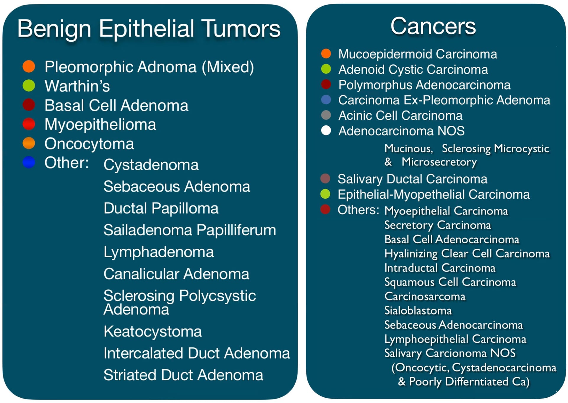
When imaging reveals a mass within the parotid or another salivary gland, a tissue sample is often the critical next step. Salivary gland biopsy performed as a fine-needle aspiration (FNA) or core needle biopsy allows the pathologist to examine the cellular architecture of the lesion and guide treatment planning: surgery or observation. There is a large variety of Salivary gland tumors. The World Health Organization recognizes over 35 distinct histological subtypes, ranging from entirely benign lesions like Warthin’s tumors to benign tumors that can transform into cancers such as pleomorphic adenoma, and finally, highly aggressive malignancies. Because clinical examination and even advanced imaging cannot reliably distinguish benign from malignant lesions in many cases, cytopathological evaluation through biopsy provides essential diagnostic clarity that directly informs the surgical approach.
Why Biopsy Salivary Glands?
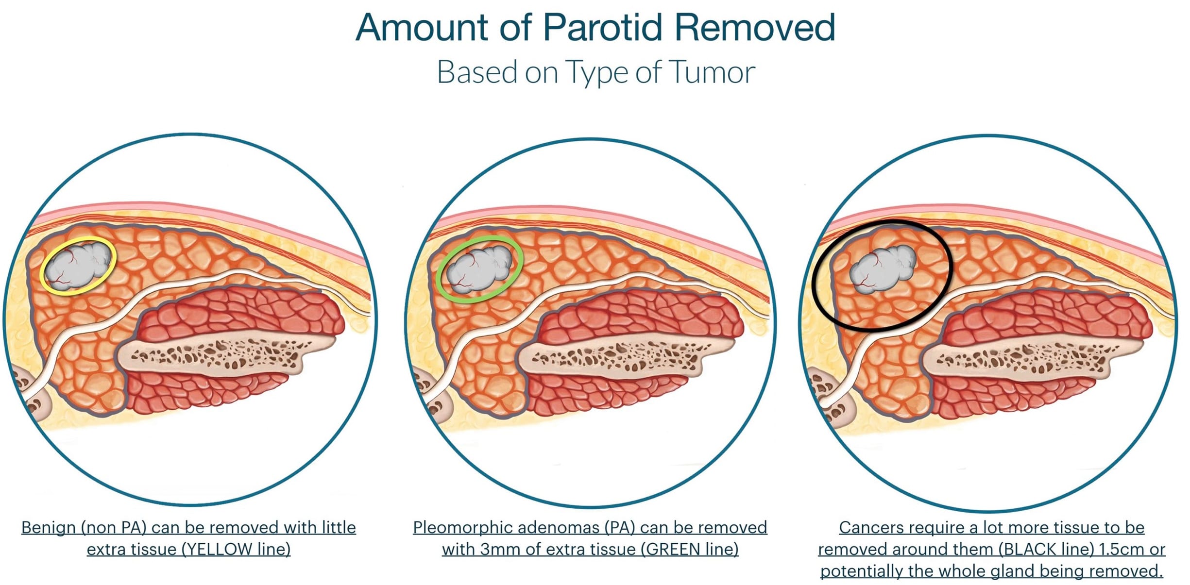
The amount of parotid tissue removed is completely related to the type of tumor present. Most benign tumors require removal of very little extra parotid tissue to assure they do not come back, but pleomorphic adenoma although benign has a higher tendency to recur so more additional parotid tissue must be removed to prevent it from recurring. Cancers require a bigger margin of safety and thus should be removed with an even larger amount of additional parotid tissue to minimize recurrence. This is very important information for your surgeon to plan your surgery.
Fine Needle Aspiration (FNA)
Fine-needle aspiration is the standard first-line biopsy technique for salivary gland masses. A thin needle (22–25 gauge) is passed into the mass — typically under real-time ultrasound guidance — and cells are aspirated for cytological analysis. The procedure takes only a few minutes, requires no incision, and can be performed in the office setting immediately following diagnostic imaging review (usually an ultrasound performed by Dr. Larian).
At the CENTER, FNA is performed by a specialist under direct ultrasound guidance, ensuring optimal needle placement and minimizing the risk of sampling error. Ultrasound guidance is especially valuable for deep-lobe lesions, small masses, or tumors with heterogeneous internal architecture where random passes may miss the most diagnostically informative regions.
The aspirated material is smeared onto glass slides and processed for cytological review. On-site rapid assessment is occasionally employed with a pathologist to confirm adequacy of the sample before the patient leaves the procedure suite, reducing the rate of non-
diagnostic specimens.
All micro-parotidectomies are combined with a reconstructive procedure to fill in the defect left behind by the removed portion of the gland. In addition to restoring symmetry to the face, this corrective procedure provides an added layer of protective tissue on top of the facial nerve, and will help prevent FREY’S SYNDROME. To achieve this, the surgeons will use muscle flaps, grafts or a combination of these to provide a positive aesthetic outcome, symmetry and ensure the patient experiences minimal to no scarring. Micro-parotidectomy is commonly performed as an outpatient procedure; most patients do not require hospitalization and can return home the same day as surgery.
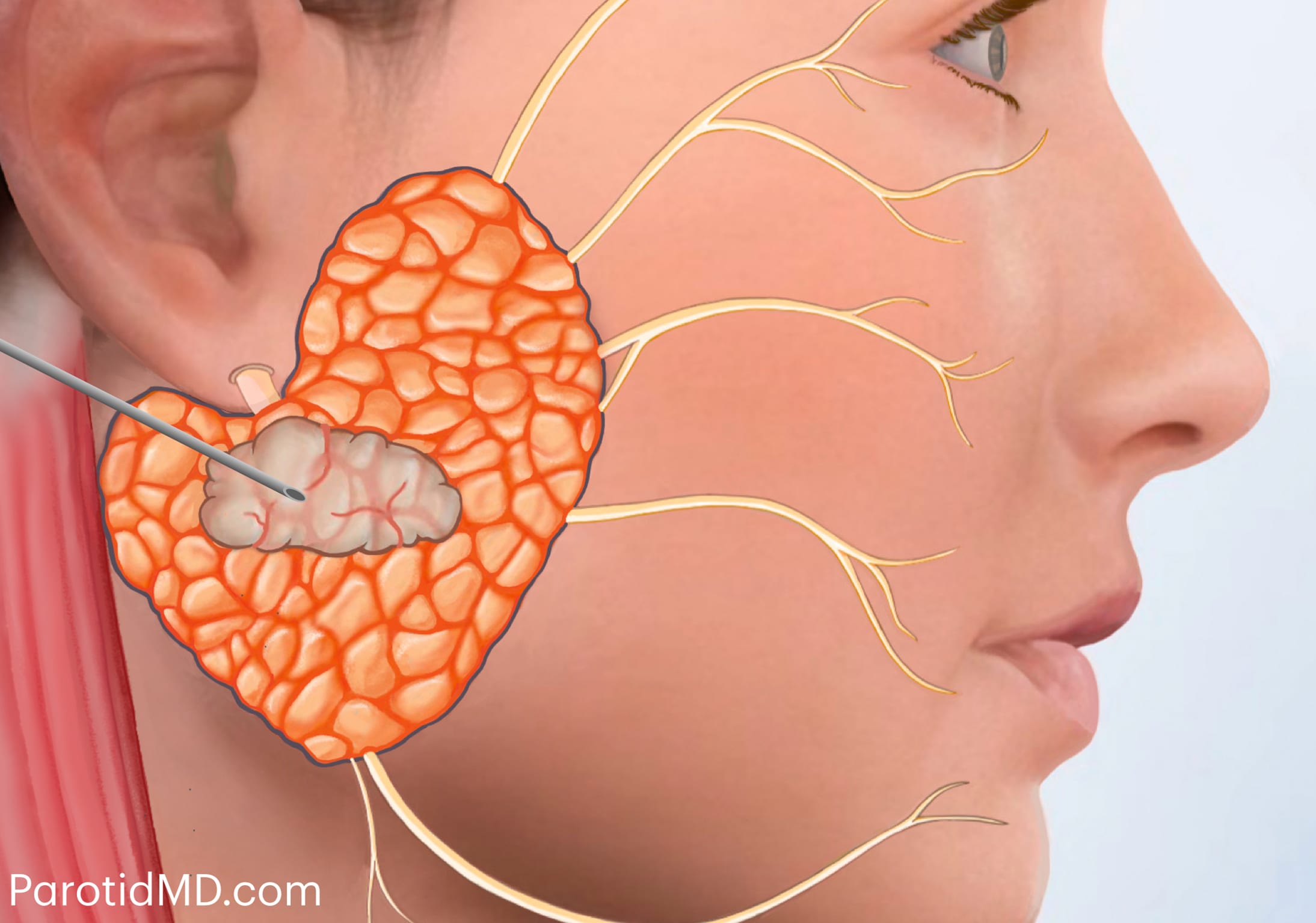
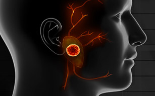
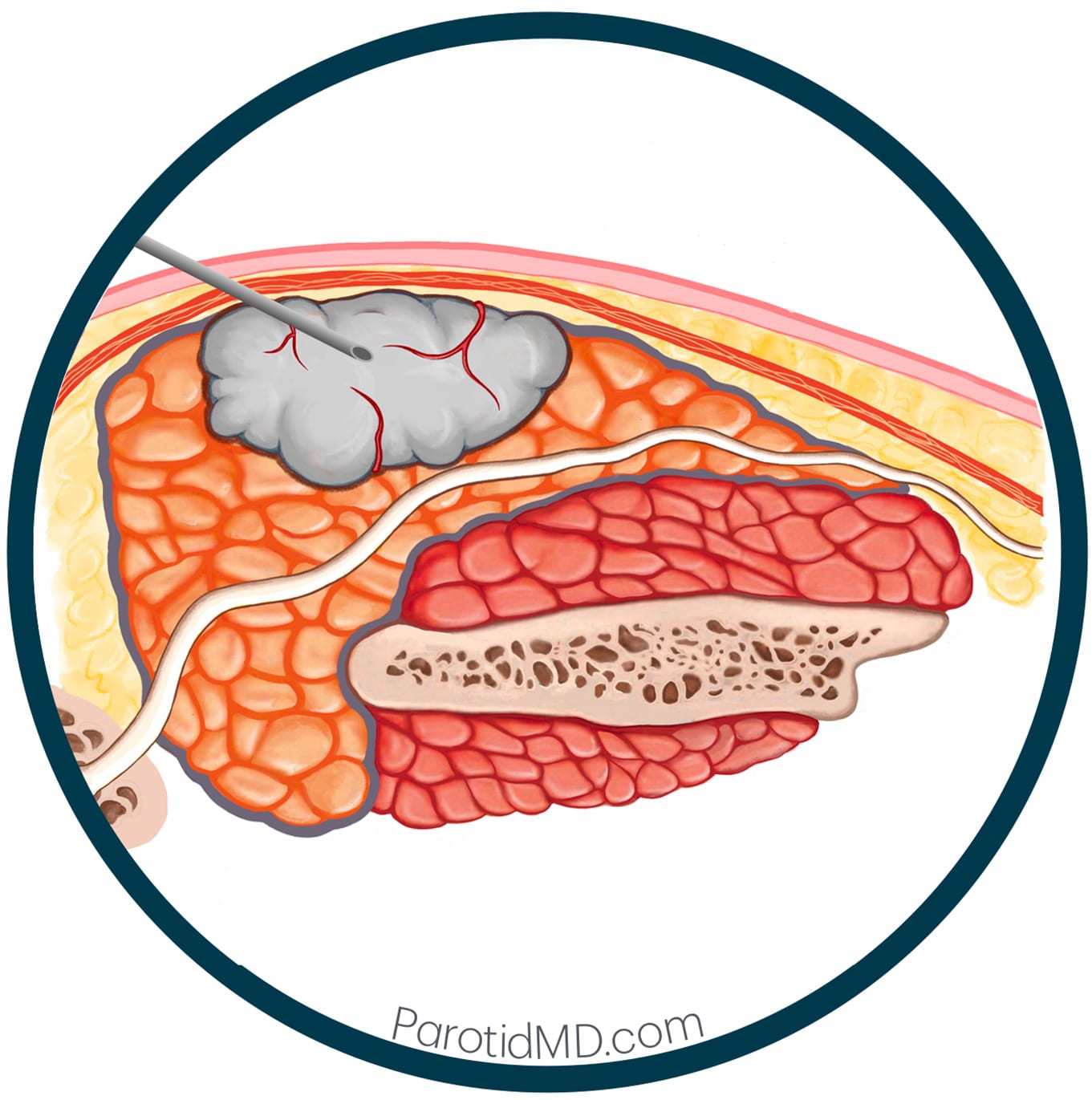
Profile & Cross-Sectional View of The Parotid Gland Tumor FNA
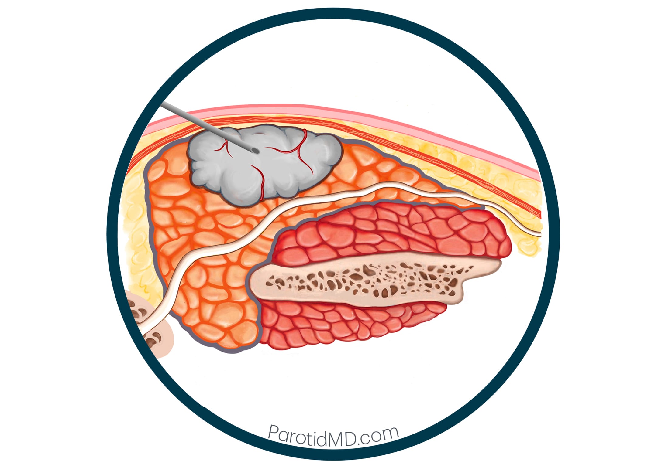
Cross-Sectional View
Under ultrasound guidance a very small needle can be safely placed inside the tumor to make sure of accuracy of the sample.
KEY ADVANTAGES
- It is safe to perform in the parotid gland without risk to the facial nerve when performed by an experienced operator.
- Using ultrasound guidance can improve accuracy and yield.
- FNA causes minimal discomfort, no requirement for incision or general anesthesia, and results that are typically available within 24–72 hours.
- Local anesthetics are used to minimize the pain & discomfort.
- You can go back to work and exercise immediately after the procedure.
DISADVANTAGES
- The more experienced the person performing the fine needle aspiration (FNA), the higher the likelihood of obtaining accurate results.
- Not every surgeon has an ultrasound machine in their office, which is the reason we at the CENTER perform our own ultrasound during your visit.
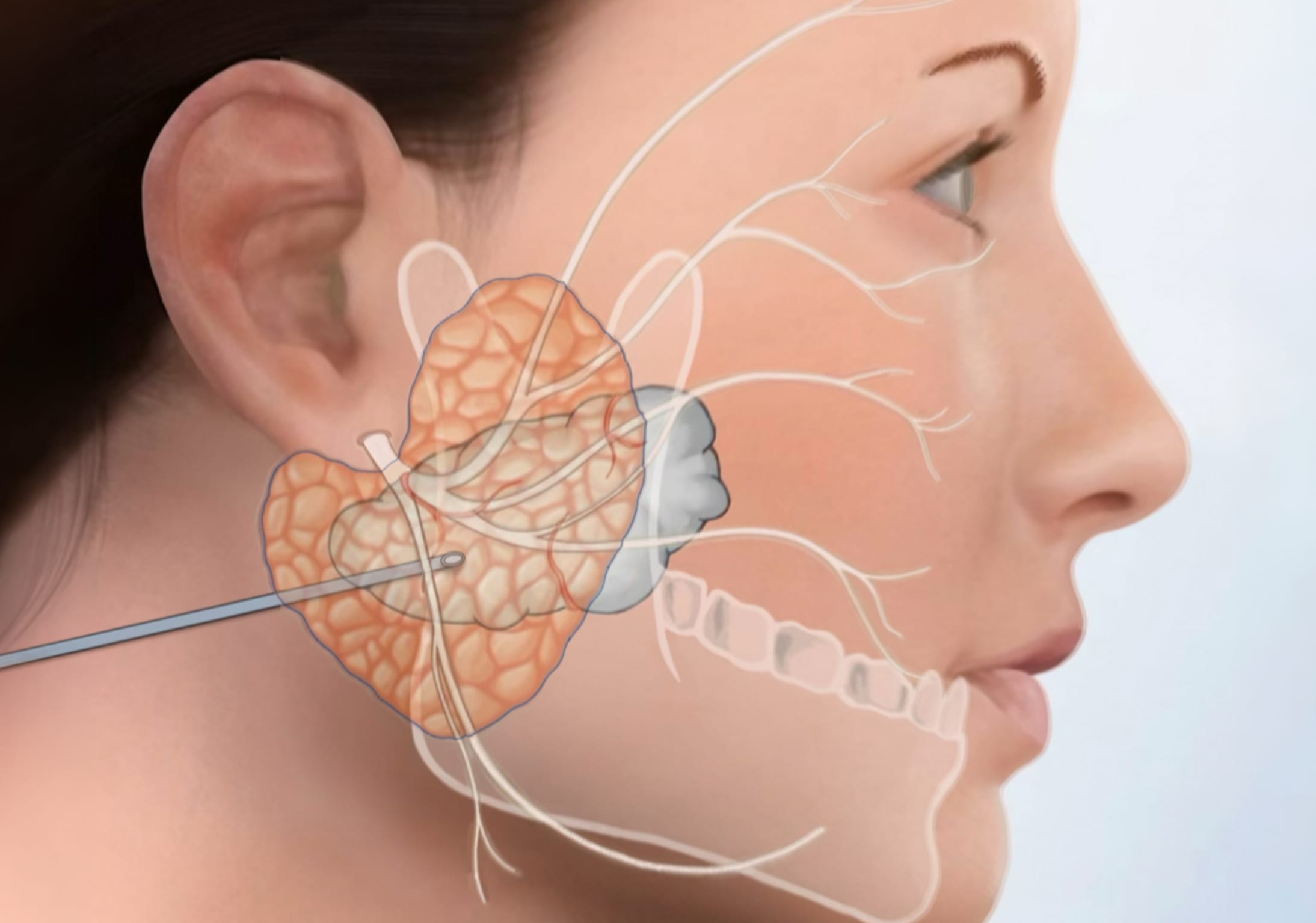
Profile & Cross-Sectional View of A Deep Lobe Parotid Gland Tumor FNA
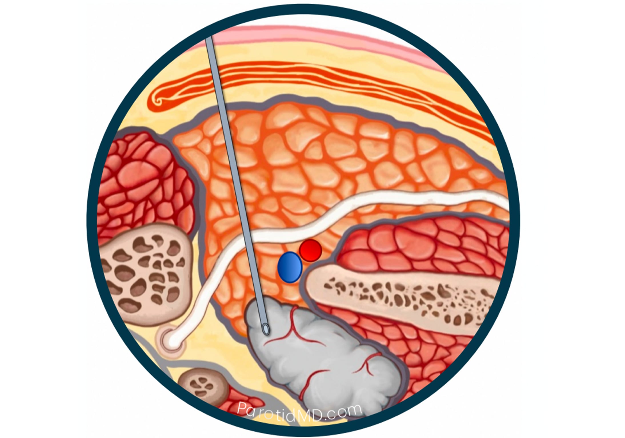
Cross-Sectional View
Under ultrasound guidance or CT guidance a very small needle can be safely placed inside the tumor to make sure of accuracy of the sample.
Deep Lobe tumors can be seen on ultrasound and an FNA can safely and easily be done with the help of the ultrasound.
Parapharyngeal Space tumors (tumors that start from the deeper most aspect of the parotid gland and grow deep to the jawbone towards the throat may not be visible on ultrasound (ultrasound cannot see through bone). In this circumstance, a CT scan can visualize the parotid tumor and an FNA can be done with CT guidance.
MILAN PATHOLOGY REPORTING SYSTEM
The Milan System for Reporting Salivary Gland Cytopathology (MSRSGC) was introduced to standardize the reporting system for salivary needle biopsies. It provides both patients and doctors with evidence-based information about the risk of cancer associated with each category of pathology report and the recommended management pathway.
The MSRSGC categorizes every salivary gland fine-needle aspiration (FNA) into one of six categories. Understanding these categories aids patients and referring clinicians in interpreting biopsy reports and comprehending the subsequent steps.
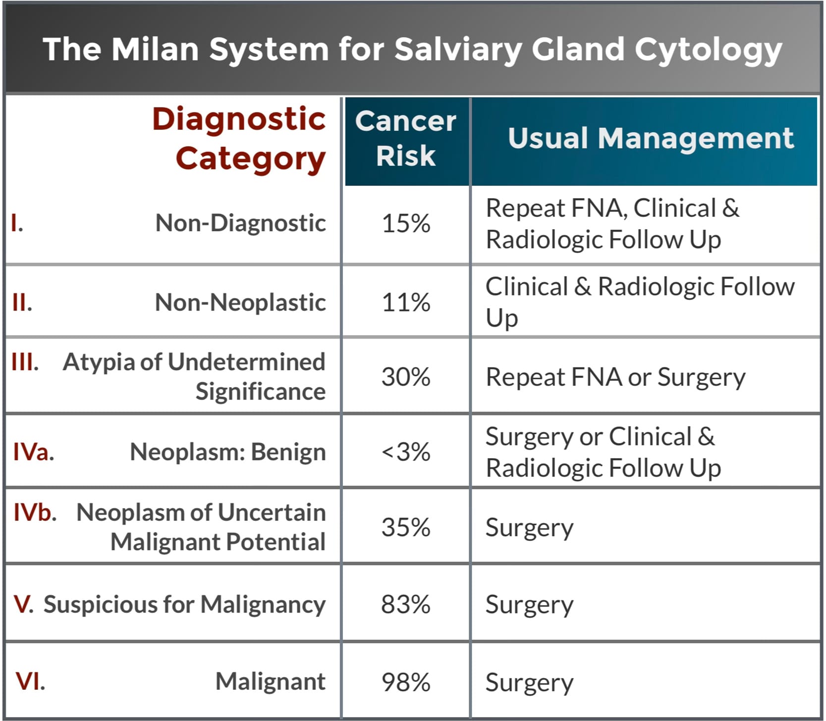
Some categories, like IVa and VI, are quite clear. For instance, when a needle biopsy reports a benign tumor, it’s accurate over 97% of the time. This means that if the biopsy indicates a Pleomorphic Adenoma (category IVa) and surgery is performed, less than 3% of the time the final pathology reveals a different, more aggressive cancer. Similarly, if a Mucoepidermoid Carcinoma (category VI) is reported, it’s extremely rare for the surgeon to discover that it wasn’t a cancer but rather something else, such as a benign tumor with unusual presentation or an inflammatory process, during or after surgery. Experienced surgeons can potentially alter these odds by analyzing scans or considering the disease’s progression, leading to a different conclusion before performing surgery, or devising alternate plans..
Other categories are less clear. Categories III and IVb carry a relatively high cancer risk, with a chance of 30% or more. In such cases, either a repeat biopsy or surgery should be performed to determine the true nature of the lesion. Your salivary gland specialist can then use the imaging characteristics, symptoms, and disease timeline to determine the best course of action.
NEEDLE BIOPSY
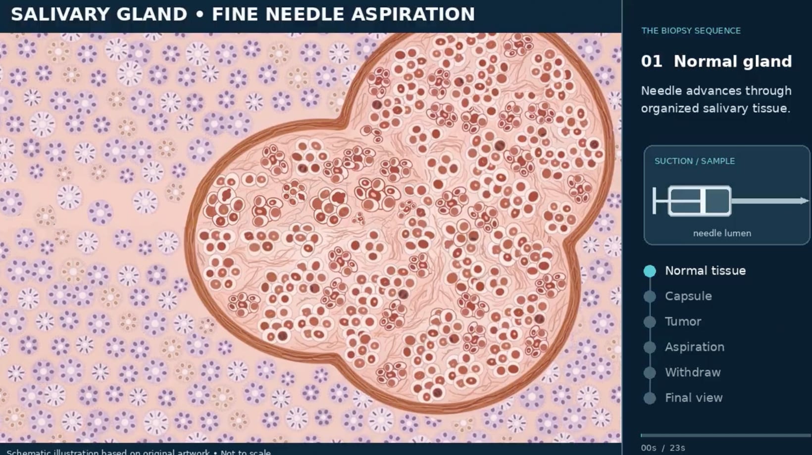
When the tumor is visible under ultrasound, a needle biopsy can be easily performed. The needle used is very thin (fine) and is inserted into the tumor. This procedure is called a Fine Needle Aspiration or FNA.
TECHNIQUE – Your doctor (a specialist conducting the needle biopsy) will use the ultrasound image to precisely locate and identify the tumor’s boundaries. The tip of a very thin needle (attached to a syringe) is clearly visible on ultrasound, so when the needle enters the tumor, it will be visible on the screen. Once the needle is inside the tumor, the plunger of the syringe is pulled back to create a vacuum, drawing the tumor cells and surrounding fibers into the hollow bore of the needle. The needle is then withdrawn, and the sample is placed on a microscope slide for examination by a pathologist to determine the type of tumor.
CORE BIOPSY
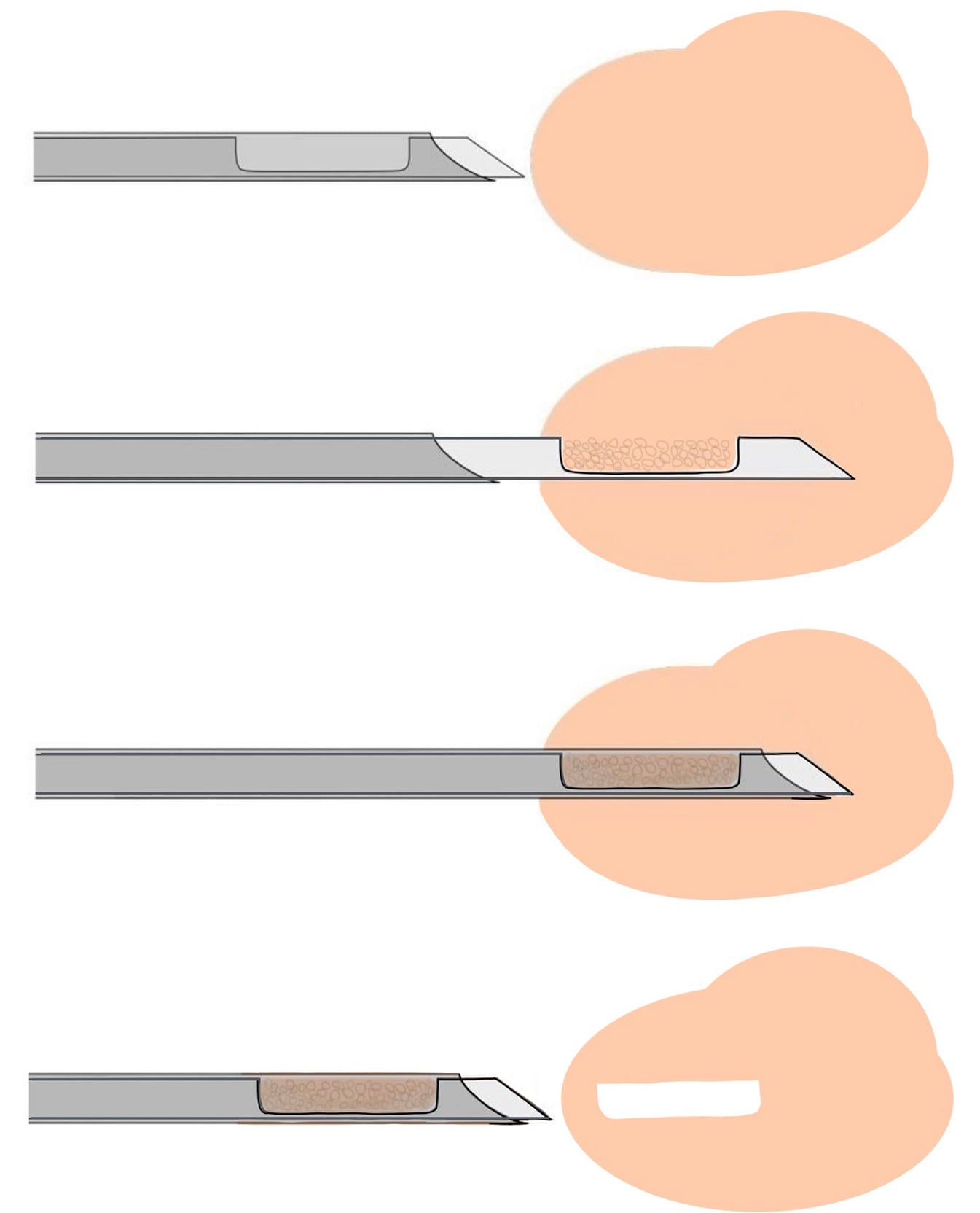
The needle used for a Fine Needle Aspiration (FNA) Biopsy is very small and thin. The cells go in the hollow bore (or hollow center) of the needle (highlighted in blue). And then are withdrawn.
A core biopsy is a larger-caliber instrument with an open side channel that captures cells (shown just below the blue highlighted needle in the diagram on the right). The technique is demonstrated below. The advantage of core biopsy is that it captures a portion of the tumor fully intact, preserving its architecture. This provides the pathologist with a greater number of cells, allowing for a more comprehensive analysis of cell formation and their relationships to surrounding tissue. Additionally, there is a larger amount of tissue available for specialized testing, which can aid in further identifying the tumor and potentially determining its molecular makeup.
A core biopsy is particularly beneficial when lymphoma is suspected. Needle biopsy alone cannot capture enough cells to accurately diagnose lymphoma. Core biopsy, on the other hand, can effectively do so.
Sometimes, when a needle biopsy doesn’t clearly reveal the type of tumor, a core biopsy can be used to obtain more tissue for better identification. A core biopsy involves a larger needle and is slightly more painful.
TUMOR SPREAD
Does needle biopsy cause the spread of tumors?
This is a common concern for both patients and doctors. Fortunately, it has been extensively studied.
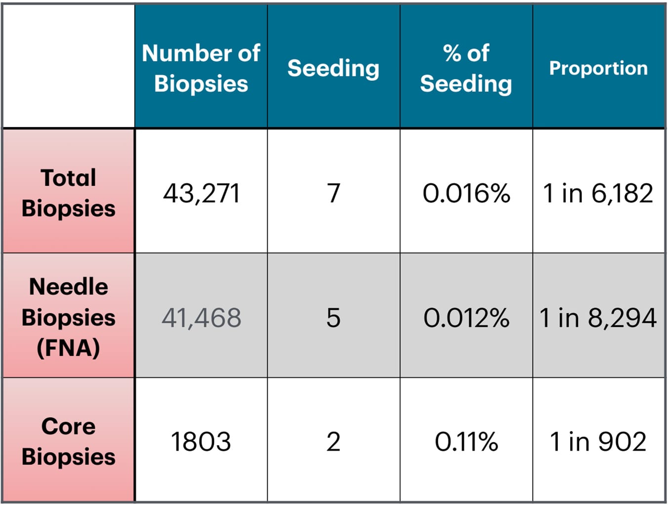
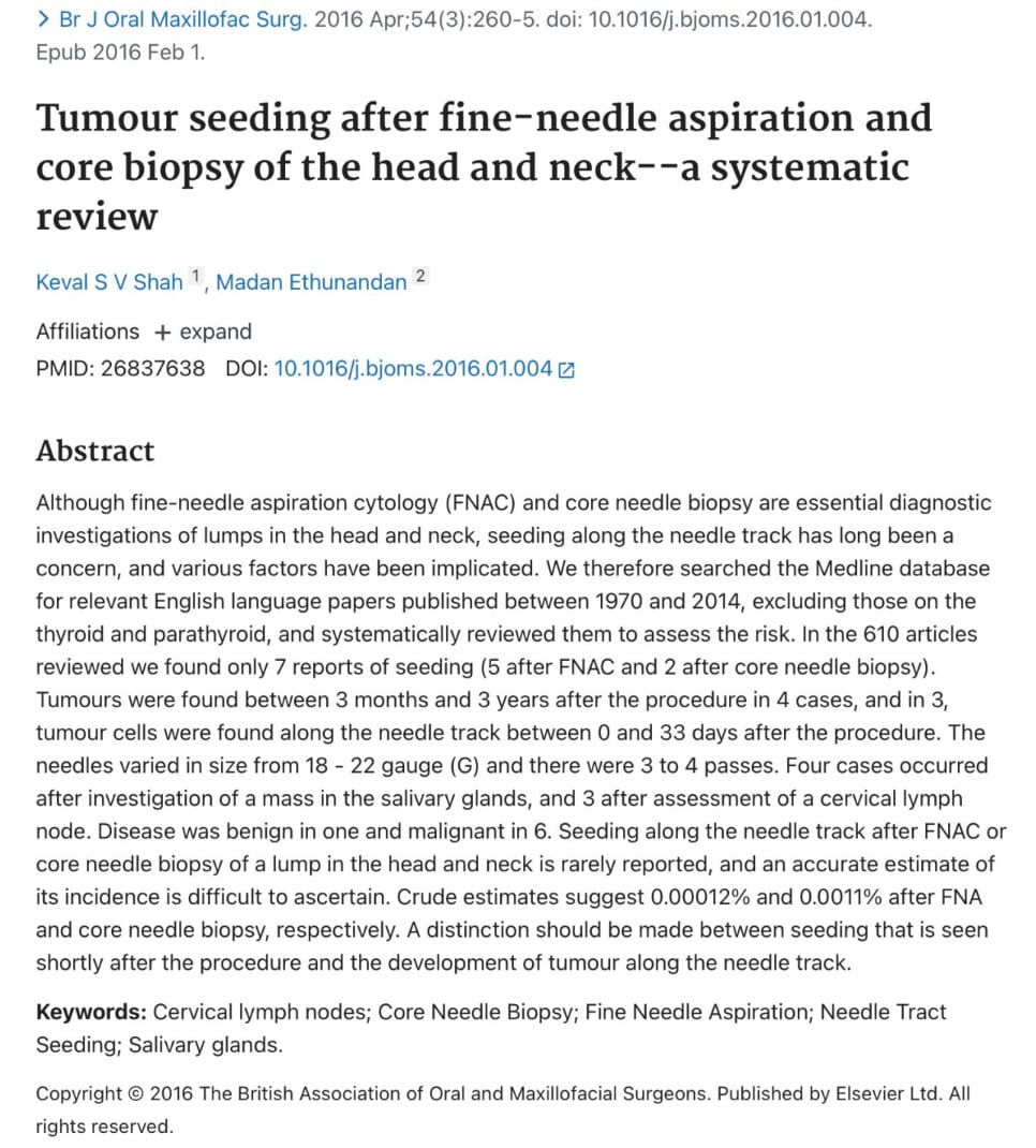
In 2016, a published scientific article analyzed 610 published articles reporting on needle biopsies performed in the head and neck region, including salivary glands and other lumps in the neck (Shah,KSV; British Journal of Oral Maxillofacial Surgery, April 2016). The study involved over 43,000 biopsies, and only 7 cases of tumor spread were identified.
The majority of these biopsies were performed using a very small needle (FNA), which carries an extremely low risk of 0.012%. This makes needle biopsies a very safe procedure.
TUMOR SPREAD
FREQUENTLY ASKED QUESTIONS
My biopsy says benign. Do I still need surgery?
Very Important Question. The answer depends on what type of benign tumor:
- Pleomorphic Adenoma: This type of tumor has a risk of transforming into an aggressive cancer that can invade the facial nerve and cause paralysis. Therefore, when a biopsy indicates a pleomorphic adenoma, it’s recommended to remove it, especially in individuals with a long life expectancy, which provides ample time for potential transformation.
- Warthin’s Tumor: This is the second most common type of tumor that occurs in the parotid glands. It’s benign and has an extremely low chance of transforming into a more aggressive tumor. Consequently, the decision to proceed with surgery will be entirely based on the tumor’s growth rate, size, and symptoms (such as pain or recurrent infections). If the tumor is small, not growing, or growing extremely slowly and not causing any symptoms, the patient has the option of monitoring it.
- The remaining types of benign tumors in the parotid would be addressed in the same manner as Warthin’s tumors.
Can the needle biopsy tell me the exact type of tumor?
It can and often does. When sufficient cells and surrounding fibers enter the hollow bore of the needle, the pathologist can thoroughly examine the tissue and provide an accurate assessment. Occasionally, they may even perform special tests, dyes, or molecular testing (if there are enough cells in the sample obtained). Unfortunately, this does not always happen, which will force your surgeon to consider the timeline history of your specific tumor, imaging results (MRI, CT, or ultrasound findings), and the types of symptoms you may have to decide on the aggressiveness of the tumor and the best next steps.
What if the needle biopsy result is non-diagnostic?
Based on the research that led to the Milan System for Salivary Needle Biopsy Reporting, when a non-diagnostic result is obtained, the probability of the mass being cancerous is approximately 15%. This is a relatively high probability. In such a scenario, your surgeon will once again need to consider the timeline history of your specific tumor, the imaging results (MRI, CT, or ultrasound findings), and the types of symptoms you may have. These factors will help your surgeon decide the next best course of action. Options include repeating the biopsy, potentially with a larger needle (core biopsy) to obtain a higher number of cells, continued monitoring, or proceeding with surgery.
Should the pathology slides obtained from a needle biopsy be sent to another pathologist for a second opinion?
Most pathologists when they encounter a pathology specimen they are unsure about will have a second pathologist look at them. This is a fairly routine practice. If still uncertain sometimes it is sent to other institutions to be looked at as well. There is very little down side to having a different pathologist look at your slides. It does take extra time, which may be worthwhile. I suggest discussing the benefits of a second opinion with your surgeon.
Why not simply surgically remove a portion of the gland (an open biopsy)?
Unfortunately, open biopsies have a high risk of seeding the tumor into the surrounding parotid or salivary gland. Therefore, open biopsies should be avoided.
Now, this is different from performing a surgical removal of the mass and surrounding salivary tissue, followed by sending it to the pathologist during the surgery. This is known as a frozen section biopsy.
Can a biopsy be performed during surgery?
Yes, it’s called a frozen section biopsy, which usually takes about 20-30 minutes. This is a valuable tool, but it has some limitations. As you recall, there are 15 types of benign and 21 types of cancers that occur in the salivary glands. Some can be easily identified under a microscope, while most require more involved testing to be clearly distinguished. Unfortunately, this isn’t possible during surgery due to time constraints. Therefore, the frozen section pathology is generally used during surgery to answer a specific question, such as whether there’s a tumor on the facial nerve, rather than determining the type of tumor.
How do I prepare, and can I drive myself home?
No preparation is required. This is a straightforward procedure, and you can drive home afterward.
What should I expect afterward, and when should I call?
You may experience slight swelling at the biopsy site. The pain is usually minimal and will subside quickly. Occasionally, the pain may persist, and applying an ice pack can provide relief. However, if the area becomes redder and more swollen within the next day or two, it could indicate an infection. In such cases, it’s advisable to contact your doctor and potentially start antibiotics.
Meet The Team
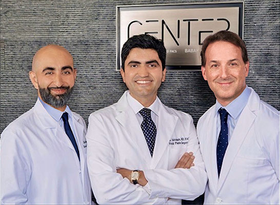
Led by board-certified parotid surgeon, Dr. Babak Larian, our team of specialists has decades of experience successfully diagnosing and treating diseases of the parotid glands with minimally invasive procedures. Distinguished by our compassionate care and cutting-edge techniques, the CENTER has developed a reputation for delivering the best parotid tumor surgery available.