There are 
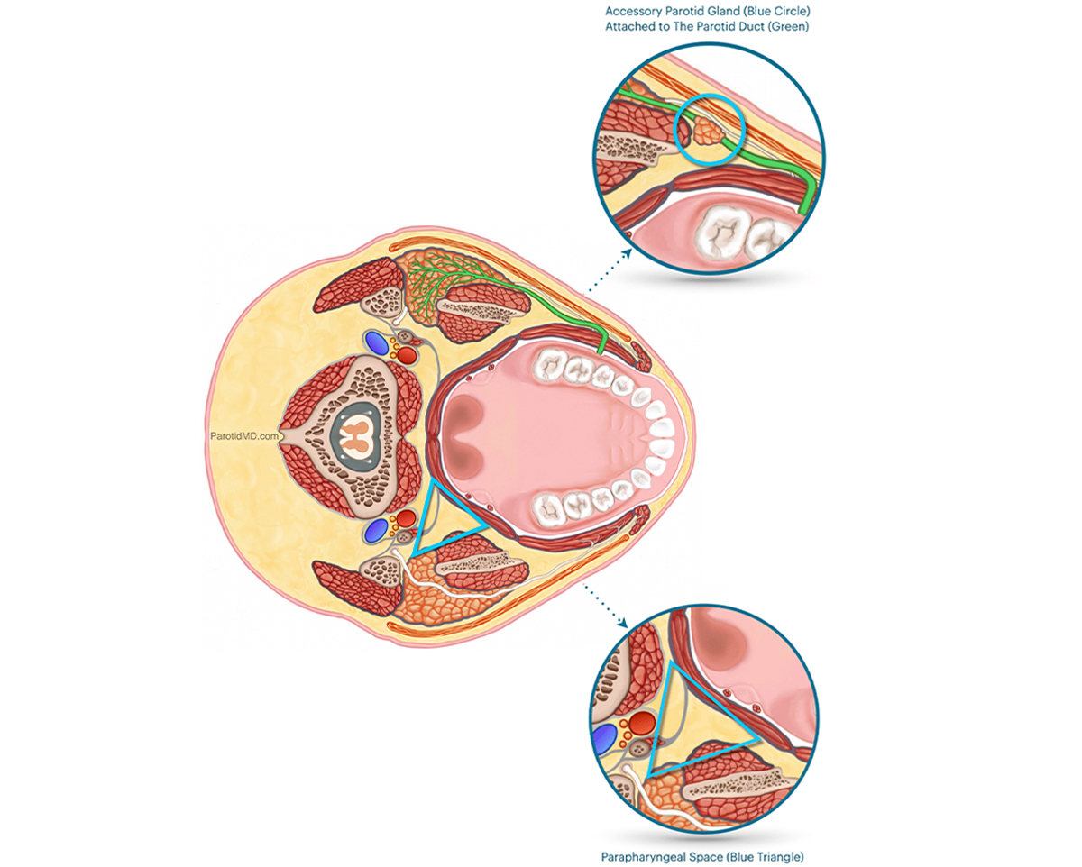
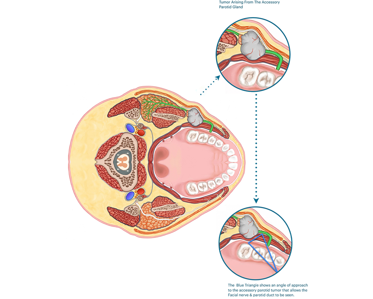
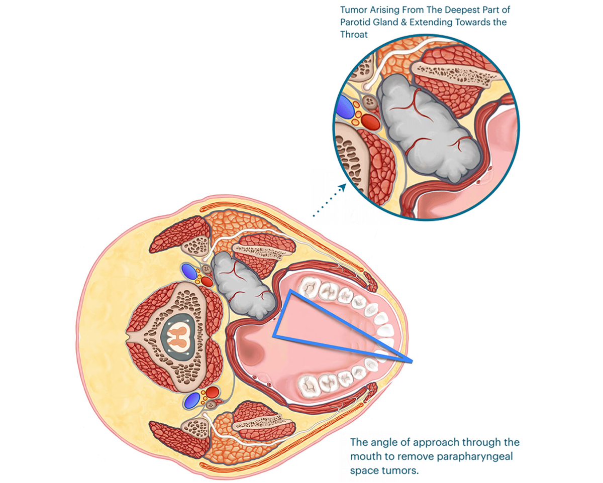
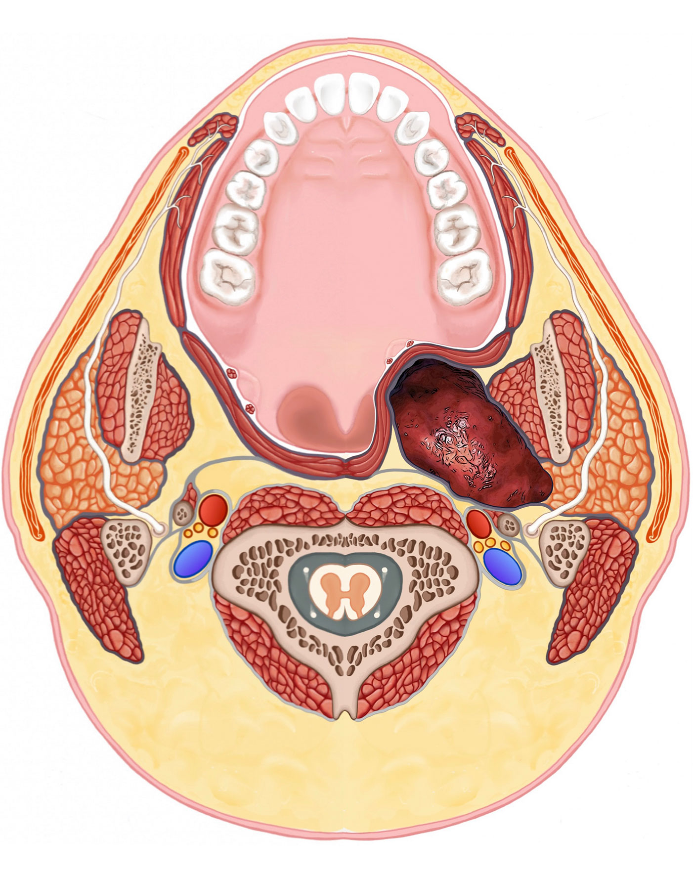
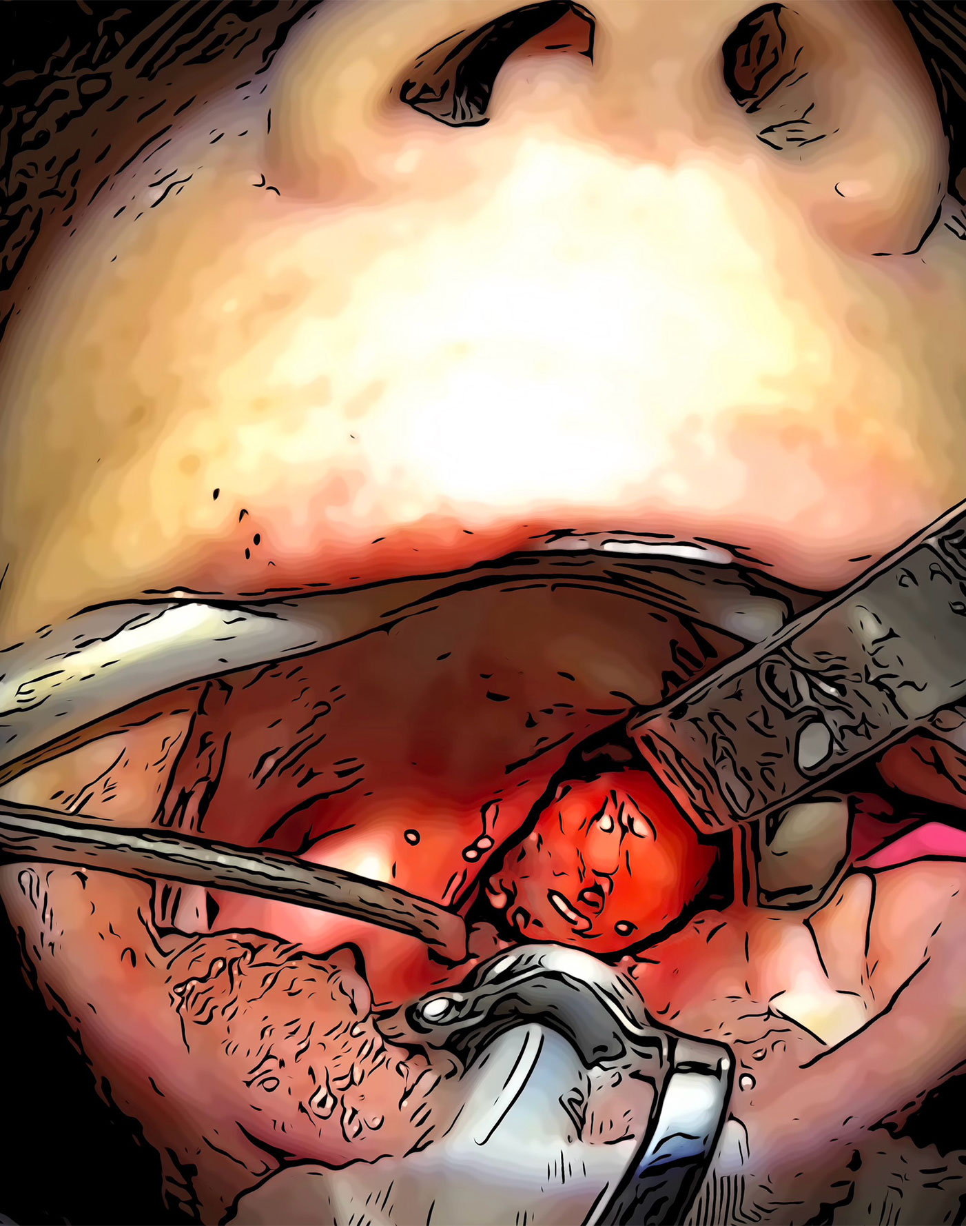
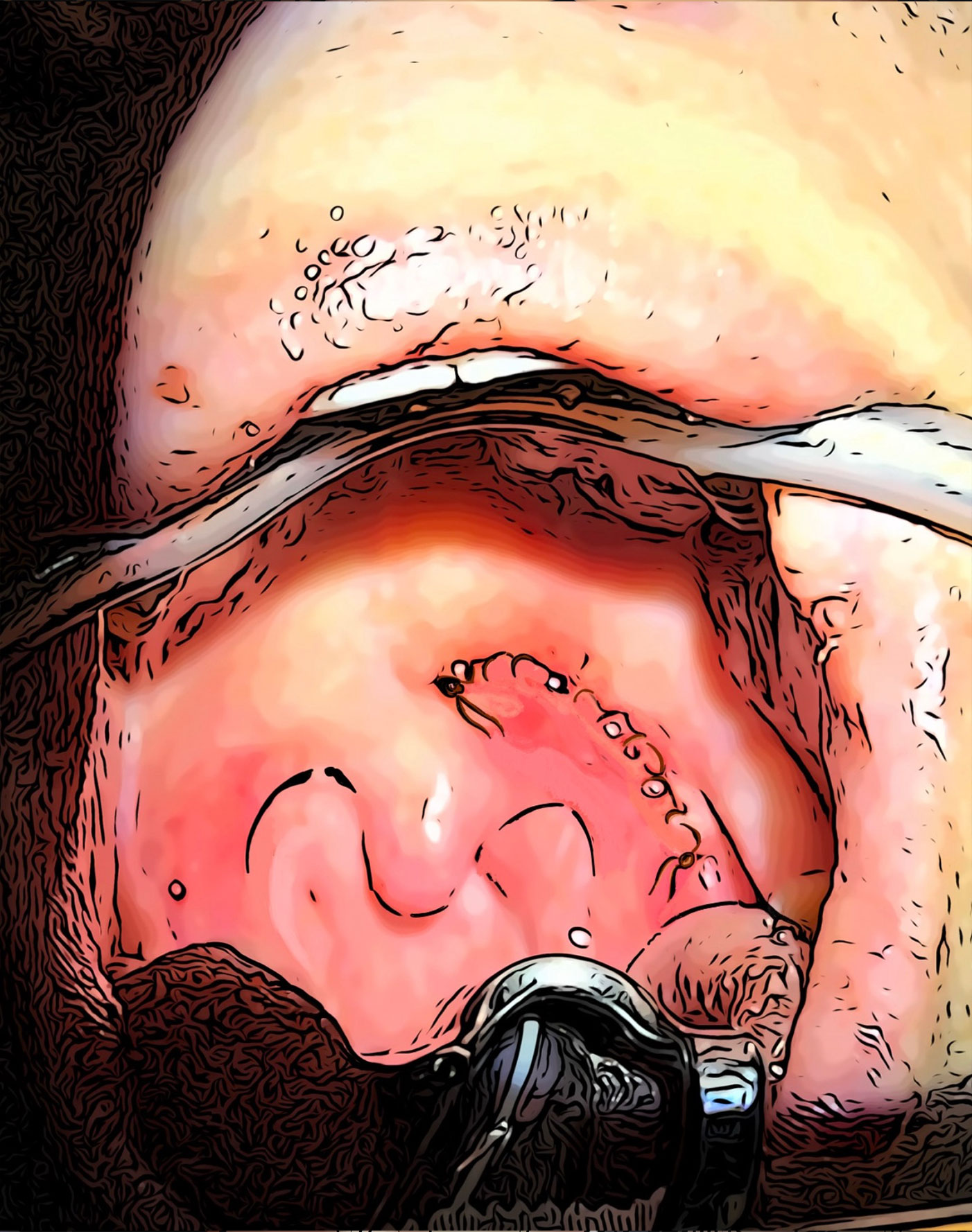
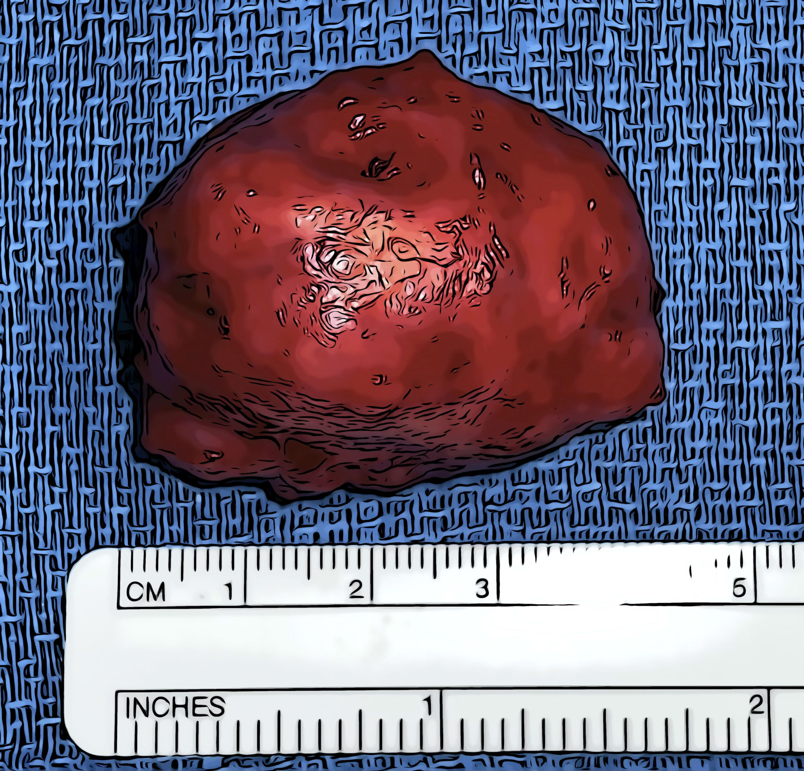
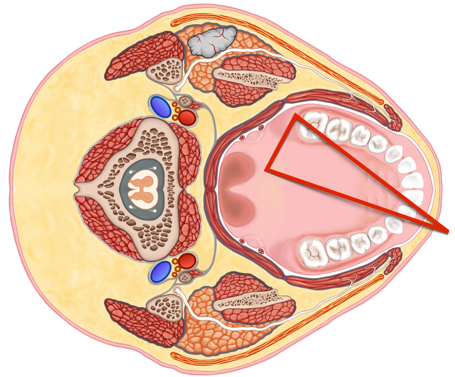
2 anatomical locations of parotid tumors that allow safe removal through mouth. In this section we’ll discuss the locations and how the surgery is done. The two locations are (1) tumors that are in the accessory parotid glands, and (2) parapharyngeal space tumors. Both of which can either be seen through the mouth or felt on examination.

The accessory parotid gland is a portion of the parotid gland that gets dragged forward by the duct (green) the develops in the gland & travels forward towards the mouth in the developing fetus. The accessory parotid glands are deep to the facial nerve and closer to the mucous membrane on the inside of the cheeks.

Accessory Parotid Gland (Blue Circle)
Attached to The Parotid Duct (Green)
Parapharyngeal Space (Blue Triangle)

The parapharyngeal space is the area deep to the parotid & jawbone that is next to and right behind the throat (pharynx). The side of this area facing the middle of the head has the styloid bone, which separates it from blood vessels & nerves. There is a barrier of tissue (called fascia – grey line) that separates this area from the middle & spine. This area is filled with mostly fatty tissue, the deep end of the parotid gland, and some lymph nodes. Because of the boundaries described above, tumors in this area generally grow in a forward direction towards the throat.

TRANS-ORAL ACCESSORY PAROTID GLAND SURGERY

The accessory parotid gland is deep to the facial nerve and closer to the mucous membrane inside the cheek/mouth. When a tumor grows in an accessory gland, it is usually deep to the facial nerve and therefore safer to access it through the mouth. Most often, if one hand is placed on the cheek skin, and the other hand’s index finger is inside the mouth, the mass can be felt between them. When detected early, these tumors tend to be mobile and can be very safely removed through the mouth. These tumors are generally found at an earlier stage because the cheek tissue is thin, and they can be easily seen or felt.

Tumor Arising From The
Accessory Parotid Gland
The Blue Triangle shows an angle of approach
to the accessory parotid tumor that allows the
Facial nerve & parotid duct to be seen.

The trans-oral approach to the accessory parotid gland tumors allows most of the surgery to be done in the area deep to the facial nerve. At the CENTER we always use facial nerve monitoring system to improve the safety to the facial nerve, as well as nerve stimulation to accurately map out the anatomy of the facial nerve in the vicinity of the tumor. Once the facial nerve branches in the area surrounding the tumor, and the parotid duct have been identified the tumor can be removed. A thorough understanding of the anatomy is crucial in being able to do this surgical approach appropriately .

TRANS-ORAL PARAPHARYNGEAL TUMOR SURGERY

The parotid gland’s deepest portion is behind and deep to the jawbone muscle. On occasion, tumors start in this area and grow inwards. This area is confined by the jawbone (to the side), spine (in the middle), and the ear bone/skull base (on the back), so the tumors that develop here can only grow in a forward direction, which is towards the throat. Once they grow large enough, they either get close to the mucous membrane of the soft palate (back of the roof of our mouth) or can push the palate, in which case we can see a bulge in the palate when we look inside the mouth.

Tumor Arising From The Deepest Part of
Parotid Gland & Extending Towards the Throat
The angle of approach through the mouth
to remove parapharyngeal space tumors.

The parotid gland’s deepest most portion is behind and deep to the jawbone muscle. On occasion tumors start in this area and grow inwards. This area is confined by the jawbone (to the side), spine (in the middle) and the ear bone & the skull (on the back), so the tumors that develop here can only grow in a forward direction which is towards the throat. Once they grow large enough they either get close to the mucous membrane of the soft palate (back of the roof of our mouth) or can push the palate forward which can be seen as a bulge in the palate when we look inside the mouth.

TRANS-ORAL PARAPHARYNGEAL TUMOR SURGERY
Live Surgery Transformed into Animation

The parapharyngeal parotid tumor is seen as a bulge in the palate on the left side. The tumor is pushing the surrounding tissue and tilting the uvula to the right. On cross-section, the tumor can be seen starting from the deep part of the parotid and extending towards the throat. This is usually seen on an MRI scan. When the limits of the tumor are visualized on the scan, then it can be estimated if the tumor size and location is appropriate for trans-oral surgery, which is the case here.

The incision (green line) is made on the palate. The tumor can be immediately seen. It is carefully separated from the surrounding fat and muscles. Once the tumor is freed on all sides, it may be removed. The video below shows the removal of the tumor through the mouth.

The incision (green line) is made on the palate. The tumor can be immediately seen. It is carefully separated from the surrounding fat and muscles. Once the tumor is freed on all sides, it may be removed. The video below shows the removal of the tumor through the mouth.

The incision is closed in multiple layers with absorbable sutures. These sutures don’t have to be later removed. The tumor is thoroughly inspected once removed. The capsule surrounding the tumor is also checked.

The tumor may be sent to the pathologist during surgery for immediate examination to see what type it is. This is done if the tumor has not been already biopsied prior to the operation, or if it has suspicious features (areas that are very hard, or has roots attaching it to the muscles, etc.). This usually takes 20 minutes, and the results are called into the operating room.

FREQUENTLY ASKED QUESTIONS

Does trans-oral parotidectomy require the person to stay in the hospital after surgery?
Fortunately, it DOES NOT. You can immediately eat after surgery; the diet is mostly liquids, smoothies, and soft foods for 5 days after surgery. The pain is minimized because the extent of surgery is much less, since you are essentially operating right on the tumor and not causing much tissue trauma to get to the tumor.

Are the facial nerves monitored during trans-oral parotidectomy?
Yes, the facial nerve monitoring system is used as an extra measure of safety for the facial nerves. In addition, when necessary, the tissue is stimulated with a physiologic nerve stimulator to see if there is a branch of the facial nerve close to the area that the surgeon is working on. It can also be used at the end of the surgery to make sure the facial nerve is fully functioning.

How can you find out if you are a candidate for trans-oral parotidectomy?
The majority of times, Dr. Larian can tell you if you are a candidate for this approach based on seeing the images of the MRI scan, which can be done remotely. Our staff can help you load the images to our software for our doctors to review them. Occasionally, Dr. Larian may need to examine you to confirm.

Why is trans-oral parotid surgery not commonly done?
It requires a thorough understanding of the anatomy, and the surgeon must be comfortable and experienced in navigating through the anatomy via the mouth. It also requires additional instruments and equipment. Mostly, it requires experience, expertise, and a team accustomed to doing this type of advanced surgery.

Why can’t all tumors in the parotid be removed via the trans-oral parotidectomy approach?
Mainly because of the facial nerve. Tumors that are inside the main parotid gland are usually in very close proximity to the facial nerve or immediately on one or a few nerve branches. As such, the surgeon would have to find the branches of the facial nerve first before removing the tumor. From inside the mouth, it is not possible to see the full facial nerve anatomy inside the parotid gland.
The approach angle through the mouth (Red Triangle) not only does not give access to that part of the gland (jawbone and muscles in the way), but would require passing through the area of the facial nerves to get to the tumor. While if this approach is from the outside, the tumor would be superficial to the facial nerve and thus much safer.

How long do out-of-town patients need to stay in town after trans-oral parotidectomy?
3 to 4 days after surgery, it would be safe to fly. All the surgical patients will have direct cellphone access to Dr. Larian and will communicate with him directly on a regular basis during the recovery period. Video follow-up visits can also be arranged for those who have extremely busy schedules.

Will Dr. Larian coordinate with our local doctors if further care is needed for the tumor?
Yes, once you are a patient at the CENTER, you will always remain a patient. Dr. Larian prefers to stay in the treatment team if further care is needed. For example, if the tumor ends up being a cancer (which happens a minority of times), Dr. Larian will want to coordinate your care with local oncologists and radiation oncologists.

Meet The Team

Led by board-certified parotid surgeon, Dr. Babak Larian, our team of specialists has decades of experience successfully diagnosing and treating diseases of the parotid glands with minimally invasive procedures. Distinguished by our compassionate care and cutting-edge techniques, the CENTER has developed a reputation for delivering the best parotid tumor surgery available.

Learn More >>

REQUEST YOUR CONSULTATION TODAY

Call us at (310) 461-0300 to schedule an appointment.
SCHEDULE A CONSULTATION