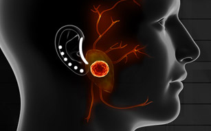
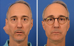
Well hidden just below the skull, deep lobe tumors are challenging to detect and pose a significant risk to delicate nerves, blood vessels and the throat. Over time, these tumors put pressure on the surrounding structure and may become cancerous. In these cases, Dr. Babak Larian and his team of specialists are able to safely remove the mass while preserving the surrounding nerves and structures.
Anatomy of The Parapharyngeal Space
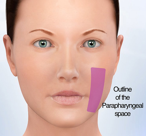
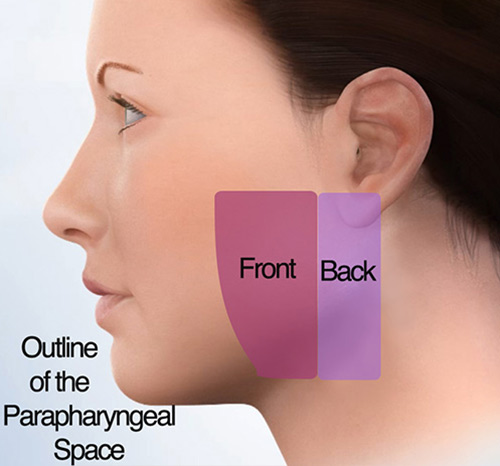
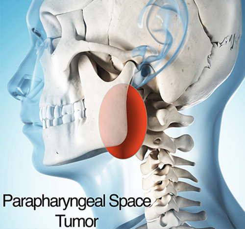
The space affected by deep lobe tumors is referred to as the parapharyngeal space, located just below the skull and extending down into the neck. The carotid artery divides the parapharyngeal space into two distinct portions - the front and back space - both of which contain many important structures.
The front part of the parapharyngeal space -- also called the Pre-Styloid Space -- contains the parotid gland, minor salivary glands, fat, and minor nerves and blood vessels. The back part of the space -- called the Post-Styloid Space -- contains the carotid artery, internal jugular vein, vagus nerve, glossopharyngeal nerve, hypoglossal nerve, sympathetic chain and lymph nodes.
An Overview of Deep Lobe Parotid Tumors
Deep lobe tumors -- also known as parapharyngeal tumors due to their location -- often present as asymptomatic masses discovered during a routine physical examination or incidental CT scan or MRI. Because the parapharyngeal space is well hidden behind the jaw, these tumors are often difficult to detect. In the event that symptoms are present, they are most often subtle and commonly due to the tumor exerting pressure on the nearby structures.
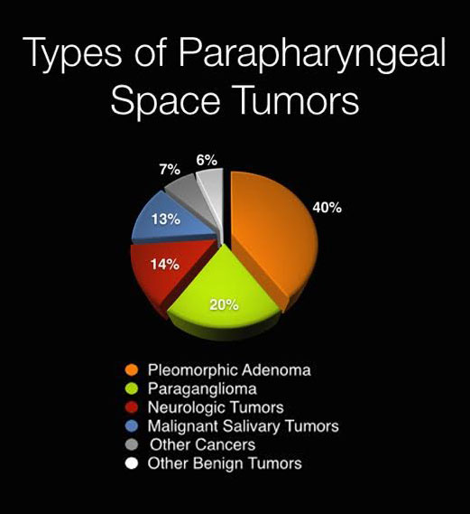
When a deep lobe tumor is located in the front portion of the parapharyngeal space, it is commonly of salivary gland origin. Although the vast majority of these tumors are Pleomorphic Adenomas, any type of salivary gland tumor or cancer can occur here.
The majority of tumors in the back portion of the space develop due to pressure and chemical receptors on the blood vessels called Paragangliomas. In these cases, most tumors are benign, but not all. Additionally, tumors in this space may also develop from the covering of nerves called schwannomas or neurofibromas, a great majority of which are also benign.
Diagnosis
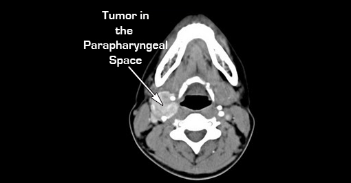
The diagnosis of a deep lobe tumor is commonly made based on MRI studies or CT scans. Because the parapharyngeal space is well hidden behind the jaw near numerous nerves and blood vessels, it is very difficult to perform a biopsy. In most cases, a fair estimation will be done based on MRI findings, but the definitive and final diagnosis is confirmed once surgery is complete.
Surgery for Deep Lobe Tumors
Deep lobe tumors typically require surgical treatment because they tend to continue growing over time, exerting pressure on the surrounding nerves, blood vessels and throat. Additionally, some benign tumors have the potential to become cancerous -- another reason surgical removal is recommended.
The team at the CENTER for Advanced Parotid & Facial Nerve Surgery is extensively experienced in the safe and successful removal of deep lobe tumors. The procedure is frequently performed through a minute incision in the neck as an out-patient procedure, except in very advanced cases.

Meet The Team
Led by board-certified parotid surgeon, Dr. Babak Larian, our team of specialists has decades of experience successfully diagnosing and treating diseases of the parotid glands with minimally invasive procedures. Distinguished by our compassionate care and cutting-edge techniques, the CENTER has developed a reputation for delivering the best parotid tumor surgery available.
Learn More >>Request your consultation today
Call us at (310) 461-0300 to schedule an appointment.
Schedule a Consultation >>


